|
|
Fasciola hepatica | ||||||||||||||||||||||||||||||||||||||||||||||||||||||||||||||||||||||||||||||||||||||||||||||||||||||||||||||||||||||||||||||
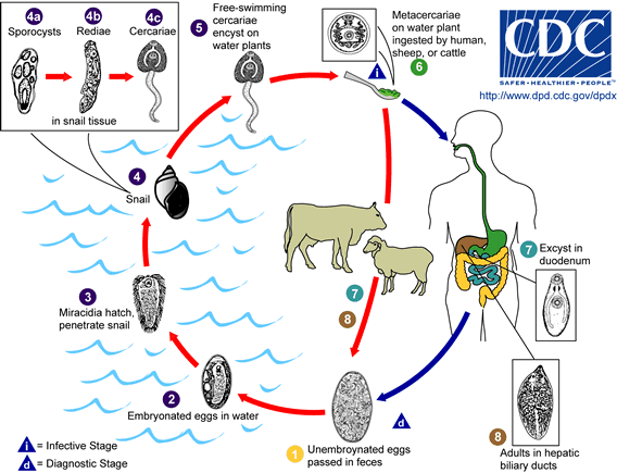
The common name of this parasite, the "sheep liver fluke," is somewhat misleading since this parasite is found in animals other than sheep (including cattle and humans), and the parasite resides in the bile ducts inside the liver rather than the liver itself. This species is a common parasite of sheep and cattle and, therefore, relatively easy to obtain. Thus, in introductory biology or zoology courses, it is often used as "THE" example of a digenetic trematode. This species has been studied extensively by parasitologists, and probably more is known about this species of digenetic trematode than any other. The adult parasites reside in the intrahepatic bile ducts, produce eggs, and the eggs are passed in the host's feces. After passing through the first intermediate host (a snail), cercariae encyst on vegetation. The definitive host is infected when it eats the contaminated vegetation. The metacercaria excysts in the definitive host's small intestine, and the immature worm penetrates the small intestine and migrates through the abdominal cavity to the host's liver. The juvenile worm penetrates and migrates through the host's liver and finally ends up in the bile ducts. The migration of the worms through the host's liver, and the presence of the worms in the bile ducts, are responsible for the pathology associated with fascioliasis. Fasciola hepatica is found in parts of the United States, as well as in Great Britian, Ireland, Europe, the Middle East, the Far East, Africa, and Australia. Fascioliasis in sheep and cattle results in animals that show low productivity (low weight gain, low milk production, etc.). Also, in many countries, livers from animals infected with F. hepatica are condemned as unsuitable for human consumption. This not only results in a significant economic loss to ranchers and farmers, but it also results in the loss of an important source of protein. The infection can be diagnosed by finding eggs in the feces of animals, but the eggs are difficult to differentiate from closely related species such as Fasciolopsis buski. Several immunological methods for diagnosis are available.
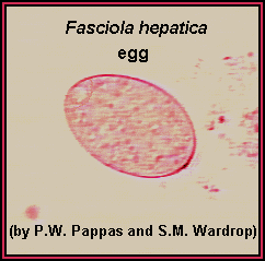
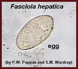
 Another example of a Fasciola hepatica egg, showing the operculum. (Original image from: The Atlas of Medical Parasitology.)
 A stained whole mount of an adult Fasciola hepatica; approximate length = 20 mm. The internal organs of this species are characteristically highly branched, thus making it very difficult to differentiate the various internal organs. Life Cycle of the Fasciola hepatica
18 day parasite cleanse by Dr. Hulda Clark. | ||||||||||||||||||||||||||||||||||||||||||||||||||||||||||||||||||||||||||||||||||||||||||||||||||||||||||||||||||||||||||||||||